While X-ray imaging is often thought of as a medical technology,
applications for it have advanced enormously during the past 100 years
of its use.
European scientists now says that a stronger type of X-ray could be
used as a new non-destructive way to the detailed chemical composition
of all kinds of objects, ranging from meteorites to fossils.
On Sunday, researchers working at the European Synchrotron Radiation
Facility (ESRF) in Grenoble, France, published a paper in the journal
Nature Materials describing a new way of rendering images of the
chemical composition of samples by using particular types of X-rays.
That means the synchrotron facility - a circular construction with an
800-meter circumference - can be used to differentiate between graphite
and diamond, for instance. Although both substances are pure carbon,
they have completely different properties as the chemical bonds between
the carbon atoms are different.
 There are only a handful of synchrotron facilities in EuropeSimo
Huotari a physics researcher at the University of Helsinki, is one of
the scientists who discovered the technique. He says it came as a "lucky
finding" while doing routine work on the ESRF facility's system of
mirrors and X-ray detectors.
There are only a handful of synchrotron facilities in EuropeSimo
Huotari a physics researcher at the University of Helsinki, is one of
the scientists who discovered the technique. He says it came as a "lucky
finding" while doing routine work on the ESRF facility's system of
mirrors and X-ray detectors.
"When the X-ray beam was going through a sample, the X-ray mirror was
taking an image of what the beam sees inside the sample, and was
reflecting that image on the detector," he told Deutsche Welle. "We were
suddenly able to start forming three-dimensional images."
Conventional X-ray images are created based on the amount of X-rays
which pass through a particular mass. Dense bones absorb more X-rays
than flesh does, making it possible to expose a contrasting image.
But the synchrotron radiation technique uses X-rays powerful enough
to kill biological entities, which is why it is never used on biological
samples. The synchrotron uses a system of curved mirrors and detectors
to measure what scatters back from a sample, rather than what passes
through it.
"If you have oxygen or you have carbon, the characteristic energy
loss is completely different," Huotari said. "So after detecting the
photon we can say whether it was deflected by an oxygen atom, or by a
carbon atom."
Highly specialized equipment
Much of what the new synchrotron radiation technique is capable of
could be done before, however, the difference is that now scientists no
longer have to destroy a sample to peer at a sample's chemical makeup.
 Battery research is extremely important to many industriesThat
means they could observe the chemical changes a functional lithium ion
battery goes through as it is charged and discharged, for instance. Or,
they could non-destructively study the chemical composition of valuable
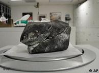
samples like meteors or fossils buried in rock.
Battery research is extremely important to many industriesThat
means they could observe the chemical changes a functional lithium ion
battery goes through as it is charged and discharged, for instance. Or,
they could non-destructively study the chemical composition of valuable
samples like meteors or fossils buried in rock.
"In a lithium battery, for example, the chemistry of lithium is
exactly the important thing that you need to look at," Huotari said.
"But so far there has been no technique to look at the chemistry of
lithium. It's a very light material, and is nearly transparent to X-rays
when it's embedded deep inside your operational battery."
Christian Schroer, a physics professor at the Dresden University of
Technology, said the new technique can render chemical properties which
could previously only be detected on surfaces or in very thin samples.
"We're now capable of really looking into objects, especially when
samples are otherwise not very accessible," he told Deutsche Welle.
But Schroer added that despite its potential, the technique is highly
specialized and therefore unlikely to be used in widespread research.
There are only a handful of synchrotron radiation facilities in Europe,
as he pointed out.
"In general you have to have a very special sample to use a synchrotron radiation facility," he said.
Visualizing a chemical environment
Christian Sternemann, a physics professor at the Dortmund University
of Technology, said one of the major advantages of the new technique is
that materials can be studied within complicated chemical surroundings.
 Geology and space exploration could also benefit"From
my point of view I think it will have a major impact on research,
because even for applications where you are looking for light elements,
it's a unique technique," he told Deutsche Welle. "I think there will be
impact especially if finer X-ray beams can be used which are highly
focused, and if the intensity one is able to bring to a sample is
optimized."
Geology and space exploration could also benefit"From
my point of view I think it will have a major impact on research,
because even for applications where you are looking for light elements,
it's a unique technique," he told Deutsche Welle. "I think there will be
impact especially if finer X-ray beams can be used which are highly
focused, and if the intensity one is able to bring to a sample is
optimized."
The University of Helsinki's Huotari also said a major advantage of
the technique is its ability it gather information about the chemical
surroundings of molecules.
"By fine-analysing the energy losses, we can also determine in what
kind of chemical environment that atom was - what kind of molecule it
was bound to, or what kind of crystal it was bound to," he added.
Author: Gerhard Schneibel
Editor: Cyrus Farivar